May 19 2009
Clitoris – Your Orgasm and Pleasure Button
Your clitoris is one of the most important parts of your sexual anatomy. Humans have sex for two reasons:
- Recreation (pleasure)
- Procreation (making babies).
Your clitoris serves absolutely no role in making babies. As such, it is not part of the reproductive system. Your clitoris has only one function: sexual pleasure. As a matter of fact, the clitoris together with her brain is at the very center of a woman’s sexual pleasure. It is the one and only organ in the entire human body, male and female, that has only one purpose – sexual pleasure and orgasm. The clitoris is endowed with the richest concentration of nerve endings (about 8,000) with the widest network of connections to other nerves in the human body. The percentage of woman who can experience sexual ecstasy and orgasm without their clitoris being a central part of it, is very small indeed. Even then it is thought that the stimulation of the internal branches of the clitoral complex might actually be involved.
The clitoris is analogous to the male penis for sexual arousal. erection and pleasure. Unlike the male penis, it does however not have any other function, or glands, or tubes running through it. It is internally divided into a left and right half (invisible from the outside, just like a penis) and it is completely sealed with no external exit or entrance. Its 8,000 nerve endings feed back to the pleasure centers of the female brain during sexual stimulation until these circuits are so overloaded that it will cause an orgasm.
As such, the female clitoris is the human female orgasm button. This is not always made clear to woman. The clitoris is the guaranteed way to orgasm. No other part of the female anatomy can make this claim. only the clitoris.
What does the clitoris look like?
Here are some medical descriptions of the clitoris and clitoral complex. If you are not interested in the medical descriptions, just skip the text on the grey background.
According to Grey’s Anatomy of the Human Body from Ed 20 (published in 1918):
“Anteriorly, each labium minus divides into two portions: the upper division passes above the clitoris to meet its fellow of the opposite side, forming a fold which overhangs the glans clitoridis, and is named the preputium clitoridis; the lower division passes beneath the clitoris and becomes united to its under surface, forming, with its fellow of the opposite side, the frenulum of the clitoris. On the opposed surfaces of the labia minora are numerous sebaceous follicles.
The Clitoris is an erectile structure, homologous with the penis. It is situated beneath the anterior labial commissure, partially hidden between the anterior ends of the labia minora. It consists of two corpora cavernosa, composed of erectile tissue enclosed in a dense layer of fibrous membrane, united together along their medial surfaces by an incomplete fibrous pectiniform septum; each corpus is connected to the rami of the pubis and ischium by a crus; the free extremity (glans clitoridis) is a small rounded tubercle, consisting of spongy erectile tissue, and highly sensitive. The clitoris is provided like the penis, with a suspensory ligament, and with two small muscles, the Ischiocavernosi, which are inserted into the crura of the clitoris.”
According to the now widely accepted new view of the clitoris from The Journal of Urology 174 (4 Pt 1): 1189–95 (October 2005). "Anatomy of the clitoris". The Journal of Urology 174 (4 Pt 1): 1189–95 by O’Connell HE, Sanjeevan KV, Hutson JM:
“The clitoris is a multiplanar structure with a broad attachment to the pubic arch and via extensive supporting tissue to the mons pubis and labia. Centrally it is attached to the urethra and vagina. Its components include the erectile bodies (paired bulbs and paired corpora, which are continuous with the crura) and the glans clitoris. The glans is a midline, densely neural, non-erectile structure that is the only external manifestation of the clitoris. All other components are composed of erectile tissue with the composition of the bulbar erectile tissue differing from that of the corpora. The clitoral and perineal neurovascular bundles are large, paired terminations of the pudendal neurovascular bundles. The clitoral neurovascular bundles ascend along the ischiopubic rami to meet each other and pass along the superior surface of the clitoral body supplying the clitoris. The neural trunks pass largely intact into the glans. These nerves are at least 2 mm in diameter even in infancy. The cavernous or autonomic neural anatomy is microscopic and difficult to define consistently. MRI complements dissection studies and clarifies the anatomy. Clitoral pharmacology and histology appears to parallel those of penile tissue, although the clinical impact is vastly different.”
The clitoris (or as it is often called, the ‘clit’) is a large organ that are mostly hidden from view. It is actually called the clitoral complex today, because it stretches backwards (posterior) underneath the labia and around the vaginal canal, it is not just the small part you can see and feel. The clitoral complex is made up of the clitoris glans (head), clitoris neck, clitoris body, clitoris legs and clitoris bulbs.
Here is the clitoris as most people will see it:
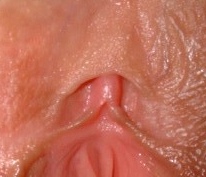
Visible Clitoris Glans peeking out from under the Clitoral Hood
(License: Lamilli [GFDL or CC-BY-SA-3.0], via Wikimedia Commons)
Here is the Clitoral Complex which is mostly hidden from view:

A picture showing the internal anatomy of the human vulva, focusing on the anatomy and location of the clitoris
(License: Amphis (Drawn by Amphis.) [Public domain], via Wikimedia Commons)
Here is what the clitoral complex looks like from the side:

Clitoral Complex viewed from the side. During arousal, excess blood supplies the clitoris’ erectile tissue causing it to swell and become erect.
(License: en:user:LearnAnatomy CC-BY-3.0 , via Wikimedia Commons)
The Clitoris was the most misunderstood and ignored part of the female anatomy for many decades/centuries. Although it is difficult to believe, the full extent of the clitoral complex was only documented in the late 1990’s by Dr. Helen O’Connell. Her research on the anatomy of the clitoris was first reported in the New Scientist magazine on 1 August 1998 (pp. 34-35) by Susan Williamson and Rachel Nowak. The scientific paper was published in the Journal of Urology in 1998 (Helen E. O’Connell, John M. Hutson, Colin R. Anderson and Robert J. Plenter, "Anatomical relationship between urethra and clitoris", Vol. 159, June 1998). In 2005, Dr. Helen O’Connel published her paper “Anatomy of the Clitoris” in the Journal of Urology. This was based on MRI studies of live and younger women, as well as the traditional anatomical dissections of human cadavers. The dissections were typically of older post-menopausal women, so it differed from the anatomy of younger women. Using the MRI the process of arousal could also be studies in a 3D plane, making it easier to see how the different parts react to arousal and making the distinction even clearer. It is quite possible that further research can discover even more of the clitoris and its function and interaction during sex.
This is just amazing to think that it took until 1998 for somebody to document the full anatomy of the clitoris. So every surgeon operating on the human female’s pelvis until then, did so without any idea of the structures they were cutting into. It was only the widespread availability of Magnetic Resonance Imaging (MRI) that made the study of the human clitoris and physiological changes during arousal and intercourse possible.
The tip (head or glans) is found at he top of the vulva where the labia majora (outer lips) and labia minora (inner lips) come together. This is at the top of the pudendal cleft (also known as the Cleft of Venus, Pudendal Fissure, Pudendal Cleavage, Pudendal Slit, Urogenital Cleft, Vulvar Slit, Rima Vulvae, or Rima Pudendi or just plain Crack or Slit in common culture). Only the glans is normally visible from the outside when the vulva it pulled open. The clitoris might also be totally invisible, even when aroused and erect. The clitoris is made from erectile tissue so it grows and become erect like a male penis when stimulated. The clitoris might be invisible when unaroused and only show its head when you get aroused. The terms ‘innie’ and ‘outie’ clitoris refers to a clitoris that are sticking out or are hidden. Both are normal.
Almost 100%, if not absolutely 100%, of woman can reach orgasm by just stimulating and vibrating the clitoris. Many women and very few men understand this fact – THE CLITORIS AND ITS STIMULATION IS WHAT CAUSE ORGASM IN A WOMAN. LITTLE ELSE, NADA, ZIP, NOTHING. This is the single most misunderstood fact, and the reason many woman are sexually unfulfilled by normal intercourse with a man. They expect the clitoris to get enough stimulation from intercourse with their male partner. The clitoris is however physically located in a place where it will not get enough stimulation to bring her to orgasm without taking special actions to bring stimulation to the clitoris.

An ‘innie’ clitoris that only gets exposed when the clitoral hood is pulled back
(License: Naumann78 (Own work) [Public domain], via Wikimedia Commons)
The well known sex therapist Betty Dobson drawing and describing the internal and external parts of the clitoris
(YouTube)
Clitoris Statistics
- The erectile structures are usually much larger in premenopausal women. This means, the size of a woman’s erectile structures are in part determined by hormone levels. An eighteen year old likely has a larger clitoris than a sixty-five year old woman.
- The urethra is surrounded on three sides by erectile tissue. There is no erectile tissue between the vagina and the urethra.
- It is perhaps inaccurate to consider the bulbs to be associated with the vestibule, as they are more closely associated with the clitoris and urethra.
- The body of the clitoris is 1 cm to 2 cm (0.4 in to 0.8 in) wide.
- The body of the clitoris is 2 cm to 4 cm (0.8 in to 1.6 in) long.
- The body and crura of the clitoris have a "deep pink" vasculature.
- The body of the clitoris projects outward from the pubic bone, versus lying against it as it is often shown.
- The crura are 5 cm to 9 cm (2.0 in to 3.5 in) long.
- The bulbs are 3 cm to 7 cm (1.2 in to 2.8 in) long.
- The bulbs have a "deep blue vasculature."
- The dorsal nerve of the clitoris is "noticeably large," being greater than 2 mm (0.08 in or 5/64th in.) in diameter. It is visible to the naked eye.
Clitoris Size
- The clitoral size was measured in 200 normal women. 99% were White; 1% were Black, 33% were using hormonal birth control.
- The women ranged in age from 14 to 56 years (mean 28, standard deviation [SD] +- 7.9, median 35.0).
- The mean transverse diameter of the glans clitoris was 3.4 mm +- 1.0 mm.
- The longitudinal diameter of the glans was 5.1 mm +- 1.4 mm.
- Total clitoral length including glans and body was 16.0 mm +- 4.3 mm (of course, now we know they were only measuring what they could see and feel.)
- The mean clitoral index was 18.5 mm^2.
- Glans length and width, clitoral index, and total clitoral length were all significantly correlated.
- Measurements of all diameters were normally distributed. Age, height, weight, or current use of hormonal birth control did not influence clitoral size.
- There was a significant difference in the glans length and width and the derived clitoral index when women that gave birth as compared with women that did not give birth yet.
- Though not statistically significant (p = 0.10), the data suggested a progressive trend toward larger total clitoral length with increasing number of offspring.
- The glans clitoris is a target organ that is responsive to androgenic stimuli and enlarges throughout life.
- The size of the glans clitoris can be quantified by determining the clitoral index (CI), which is the product of the sagittal and transverse diameters of the glans.
- 410 patients, ranging in age from 17 to 35 years, were examined.
- 95% of 249 normal women had a CI less than 35 mm^2.
- Of 85 patients with an enlarged clitoris (clitorimegaly) (CI greater than 35 mm^2) in addition to at least 1 other clinical sign of excess androgenic stimulation, 53 (62%) had abnormally high values for either or both total serum testosterone and 17-ketosteroid levels.
Clitorimegaly
Clitorimegaly is the abnormal enlargement of the clitoris. This should not be confused with the normal process of erection of the clitoris during female arousal. Clitorimegaly is defined as a clitoris at least twice the size of the average clitoris.

Enlarged Clitoris in a Healthy 22 Year Old Woman
(License: CC-BY-2.0, via Wikimedia Commons from Copcu, E, Aktas, A, Sivrioglu, N, Copcu, O, Oztan, Y. Idiopathic isolated clitoromegaly: A report of two cases. Reproductive Health. 1, 4. 2004)
Clitorimegaly can occur for 4 reasons:
- Hormonal imbalance during embryonic development causing the birth of an intersex baby or a baby with ambiguous genitalia which are not clearly male or female.
- Medical conditions in adult women like PCOS (PolyCystic Ovarian Syndrome ) or diseases affecting he ovaries and other endocrine glands. PCOS cause abnormalities in the metabolism and control of the production of androgens in the woman’s body. This does not occur in all PCOS sufferers and a particular woman might have normal androgen levels.
- Taking performance enhancing drugs like anabolic steroids, testosterone and androgens. Although illegal, this is particularly common in strength based female sports like weight lifting and body building. During sexual reassignment (female to male or FtM) androgens are used to enlarge the clitoris to form a penis.
- Taking medicines containing testosterone for medical reasons like low libido, averting osteoporosis, or as part of an anti-depressant regimen. The dose used to treat these conditions are however so low that it should not cause any significant clitoral enlargement.
References
“Clitoral size in normal women” – Verkauf BS; von Thron J; O’Brien WF, OBSTETRICS AND GYNECOLOGY. 1992 Jul;80(1):41-4.
”The clitoral index: a bioassay of androgenic stimulation.” Tagatz GE, Kopher RA, Nagel TC, Okagaki T.
“Time for rethink on the clitoris” – BBC News by Sharon Mascall 11 June 2006
“Clitorimegali” – WikiPedia
“Polycystic Ovarian Syndrome” – eMedicine from WebMD
"Female Sexual Anatomy: Clitoral & Labial Size" – TheClitoris.com
“Clitoral Hood” – WikiPedia
“Smegma” – WikiPedia
“The clitoral complex: a dynamic sonographic study.” Foldes P, Buisson O. – J Sex Med. 2009 May;6(5):1223-31. Review.
“Sonography of the clitoris.” Buisson O, Foldes P, Paniel BJ. – J Sex Med. 2008 Feb;5(2):413-7. Epub 2008 Jan 2.
“Clitoral anatomy in nulliparous, healthy, premenopausal volunteers using unenhanced magnetic resonance imaging.” O’Connell HE, DeLancey JO. – J Urol. 2005 Jun;173(6):2060-3.
“Magnetic resonance imaging anatomy of the female genitalia in premenopausal and postmenopausal women.” Suh DD, Yang CC, Cao Y, Garland PA, Maravilla KR. – J Urol. 2003 Jul;170(1):138-44.
“MRI of female genital and pelvic organs during sexual arousal.” Suh DD, Yang CC, Cao Y, Heiman JR, Garland PA, Maravilla KR. – J Psychosom Obstet Gynaecol. 2004 Jun;25(2):153-62.
“Female genitalia: dynamic MR imaging with use of MS-325 initial experiences evaluating female sexual response.” Deliganis AV, Maravilla KR, Heiman JR, Carter WO, Garland PA, Peterson BT, Hackbert L, Cao Y, Weisskoff RM. – Radiology. 2002 Dec;225(3):791-9.
“Genital and subjective sexual arousal in postmenopausal women: influence of laboratory-induced hyperventilation.” Brotto LA, Gorzalka BB. – J Sex Marital Ther. 2002;28 Suppl 1:39-53.
“Sexually responsive vascular tissue of the vulva.” Yang CC, Cold CJ, Yilmaz U, Maravilla KR. – BJU Int. 2006 Apr;97(4):766-72.
“Serial MR imaging with MS-325 for evaluating female sexual arousal response: determination of intrasubject reproducibility.” Maravilla KR, Cao Y, Heiman JR, Garland PA, Peterson BT, Carter WO, Weisskoff RM. – J Magn Reson Imaging. 2003 Aug;18(2):216-24.
“Anatomical relationship between urethra and clitoris.” O’Connell HE, Hutson JM, Anderson CR, Plenter RJ. – J Urol. 1998 Jun;159(6):1892-7.
“The anatomy of the distal vagina: towards unity.” O’Connell HE, Eizenberg N, Rahman M, Cleeve J. – J Sex Med. 2008 Aug;5(8):1883-91. Epub 2008 Jun 28. Review. Erratum in: J Sex Med. 2008 Oct;5(10):2477-9.
“Anatomy of the clitoris.” O’Connell HE, Sanjeevan KV, Hutson JM. – J Urol. 2005 Oct;174(4 Pt 1):1189-95. Review.
“Anatomical relationship between urethra and clitoris.” O’Connell HE, Hutson JM, Anderson CR, Plenter RJ. – J Urol. 1998 Jun;159(6):1892-7.
Comments Off on Clitoris – Your Orgasm and Pleasure Button